Mouth ulcers, white or red patches: get them checked
Hours: Mon 4–9 PM, Tue–Sun 9:30 AM–9 PM
2nd Floor, Horamavu Agara Road, near Nayara Petrol Bunk, Hennur Bande. Lift access available. Parking available for 4-wheelers and 2-wheelers.
Get persistent sores, painful canker ulcers, unusual white or red mucosal patches, and chronic burning mouth sensations evaluated by visiting MDS oral medicine specialists at Aureva Dental in Hennur. An ulcer that has not healed in two weeks should be examined.
Who needs an oral mucosal examination?
The soft tissue lining of your mouth responds quickly to infection, trauma, nutritional deficiencies, and tobacco exposure. While minor sores are common, persistent changes should never be ignored. We provide careful, unhurried soft tissue assessments in a calm setting.
Ulcers Lasting Longer Than 2 Weeks
An ulcer that has not healed in two weeks should be examined. Non-healing ulcers require clinical inspection to rule out chronic traumatic irritation, underlying systemic infection, or early cellular changes.
White or Red Patches
White patches (leukoplakia) or red velvety patches (erythroplakia) on the tongue, floor of the mouth, or inner cheek require prompt clinical review and regular monitoring.
Tobacco and Gutka Users
Anyone with current or past habits of chewing gutka, pan masala, betel nut, or smoking cigarettes and bidis should undergo routine oral mucosal cancer screenings every 6 months.
Burning Mouth and Recurrent Sores
Persistent burning sensations, intolerance to spicy foods, or multiple painful aphthous ulcers occurring in crops can point to oral lichen planus, geographic tongue, or systemic nutritional deficiencies that benefit from specialist care.
Traumatic Irritation from Broken Teeth
A fractured tooth cusp, worn-down filling, or misaligned orthodontic bracket can repeatedly scrape against cheek mucosa or the lateral tongue border, creating a chronic sore that will not resolve until the rough dental surface is smoothed.
Comprehensive oral medicine evaluation
1. Clinical Oral Soft Tissue Examination
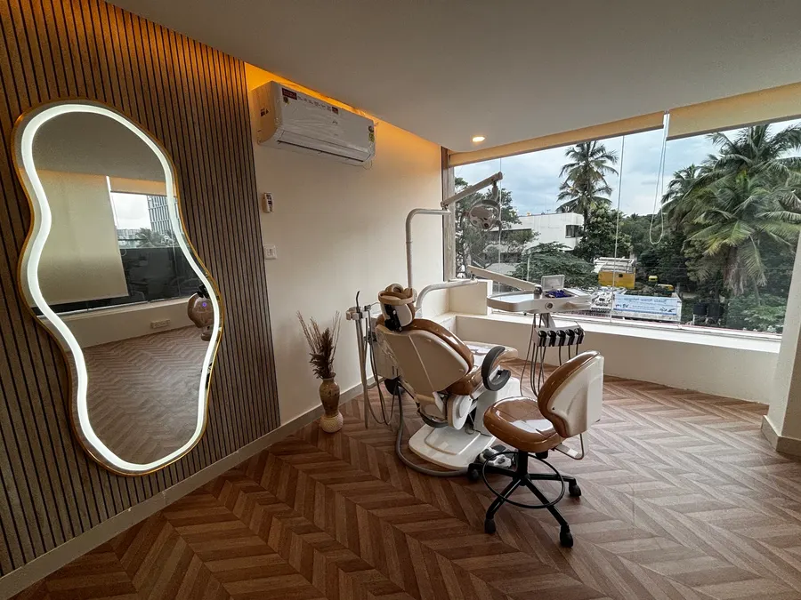
Our dentists and visiting MDS oral medicine specialists thoroughly examine your entire oral cavity. We check your inner cheeks, tongue borders, floor of the mouth, soft palate, and gums under bright LED operatory illumination. We inspect for sharp cusps, broken fillings, or rough dental crowns that may be scraping delicate mucosal tissue.
2. Oral Cancer Screening for High-Risk Groups
For individuals who consume tobacco or betel nut, we provide dedicated screening using visual inspection, palpation of neck lymph nodes, and soft-tissue texture mapping. Detecting mucosal changes early is crucial for effective intervention and health preservation.
3. In-Clinic Diagnostic Biopsy Service
When a lesion does not heal following elimination of local irritation, our visiting specialist performs a gentle incisional or punch biopsy under local anaesthetic. The procedure uses autoclaved sterile instruments and single-use sealed kits. The tissue specimen is placed in formal-saline preservative and dispatched to an accredited oral pathology laboratory for histopathological reporting.
4. Medical Management & Nutritional Guidance
Depending on diagnosis, treatment may include topical corticosteroid mucosal pastes, antimicrobial mouth rinses, barrier gels, and targeted vitamin supplementation for iron, vitamin B12, or folate deficiencies.
5. Gentle Homecare and Oral Hygiene Advice
During acute flare-ups, avoid citrus fruits, vinegar, strong spicy curries, and rough crunchy snacks that physically irritate raw ulcerated mucosa. Rinse with lukewarm salt water or prescribed chlorhexidine rinses. Use an ultra-soft bristle toothbrush with non-SLS (sodium lauryl sulfate) toothpaste to prevent exacerbating sensitive soft tissues.
How oral lesion evaluation works at Aureva
Visual Assessment
Thorough examination of oral mucosa, recording size, duration, color, and location of any ulcer or patch.
Identify Irritants
Detecting sharp enamel edges, rough restorations, or mechanical denture pressure points that can be smoothed immediately.
Medical Care / Biopsy
Prescribing topical soothing regimens or performing a minor biopsy under local anaesthetic if the lesion has persisted over 2 weeks.
Review & Follow-up
Scheduled re-evaluation 10 to 14 days later to confirm healing progress or discuss official laboratory pathology findings.
Mouth ulcer and lesion consultation fees
Initial consultation includes digital RVG X-ray. Specialised tests and biopsy procedures are quoted clearly after examination.
| Service | Fee |
|---|---|
| Clinical Oral Medicine Consultation | ₹600 |
| Specialist Oral Lesion Assessment | Price after clinic examination |
| Diagnostic Biopsy (Sample Sent to Lab) | Price after clinic examination |
| Oral Cancer Screening for Tobacco Users | Price after clinic examination |
| Topical Ulcer Care & Prescription Regimen | Price after clinic examination |
An ulcer that has not healed in two weeks should be examined. Explore all treatment fees across specialties on our clinic prices directory.
Frequently asked questions about mouth ulcers
When should I see a dental specialist for a mouth ulcer?
Most simple minor canker sores (aphthous ulcers) heal naturally within 7 to 10 days. However, an ulcer that has not healed in two weeks should be examined. You should also seek clinical review if ulcers recur frequently, cause difficulty swallowing, or appear alongside white or red velvety patches on your cheeks, tongue, or gums.
What causes recurring mouth ulcers?
Recurring mouth ulcers can stem from several factors, including accidental cheek biting, sharp edges from broken teeth or rough fillings, nutritional deficiencies (such as iron, ferritin, vitamin B12, or folate), high systemic stress, and certain food sensitivities. Our visiting MDS oral medicine specialist conducts a thorough oral examination to identify underlying local and systemic triggers.
What are white patches (leukoplakia) and red patches in the mouth?
White patches that cannot be wiped away with gauze are known clinically as leukoplakia, while persistent red velvety areas are called erythroplakia. These lesions represent changes in the mucosal lining and require professional clinical evaluation, particularly for individuals with a history of smoking, bidi, gutka, pan masala, or betel nut chewing.
How is an oral mucosal biopsy performed at Aureva Dental?
When an oral ulcer, white patch, or lump does not resolve with conservative topical care, a biopsy may be advised. The area is completely numbed with local anaesthetic so you feel only gentle pressure. A small representative tissue sample is carefully taken, autoclaved sterile instruments are used, and the sample is sent to an accredited pathology laboratory for microscopic analysis.
Can sharp teeth or ill-fitting dentures trigger persistent mouth ulcers?
Yes. Chronic mechanical irritation from a fractured cusp, chipped enamel, sharp tartar deposits, or an unstable denture border can cause persistent traumatic ulcers. Smoothing the sharp edge or adjusting the denture border often allows the tissue to heal promptly within days.
Visit Aureva Dental in Hennur Bande
Conveniently located on Horamavu Agara Road near Nayara Petrol Bunk, Hennur Bande. Open Monday 4–9 PM, Tuesday to Sunday 9:30 AM–9 PM. Lift access available. Parking available for 4-wheelers and 2-wheelers.
Book your consultation at Aureva Dental
Located on Horamavu Agara Road near Nayara Petrol Bunk, Hennur Bande. Open Monday 4–9 PM, Tuesday to Sunday 9:30 AM–9 PM.